Medical Care (Sputum Suctioning and Tube Feeding)
医療的ケア
National Exam for Certified Care Workers 介護福祉士 · 介護福祉士国家試験(筆記) · Written by Tokutei Pass based on the official study texts; not copied verbatim.
医療的ケアの法制度と特定行為の5つの範囲・医行為との境界
Legal System of Medical Care, the 5 Permitted Specific Acts, and Boundaries with Medical Practice
「喀痰吸引」や「経管栄養」は本来、医師や看護師が行う「医行為(医療行為)」ですが、2012年(平成24年)4月の改正社会福祉士及び介護福祉士法施行により、所定の研修・実地研修を修了した介護福祉士等が一定条件のもとで実施できるようになりました。
Sputum suctioning and tube feeding are inherently Medical Acts performed by physicians or nurses; however, following the April 2012 (Heisei 24) enforcement of the amended Certified Social Worker and Certified Care Worker Act, care workers who have completed prescribed training and practical training may perform them under specific conditions.
介護福祉士等が実施できる「特定行為(5行為)」は、①口腔内の喀痰吸引、②鼻腔内の喀痰吸引、③気管カニューレ内部の喀痰吸引、④胃ろう又は腸ろうによる経管栄養、⑤経鼻経管栄養の5つに限られます。
The Specific Acts (5 acts) that Certified Care Workers may perform are strictly limited to: (1) Oral sputum suctioning, (2) Nasal sputum suctioning, (3) Tracheal cannula internal sputum suctioning, (4) Tube feeding via gastrostomy or enterostomy, and (5) Nasogastric tube feeding.
これらを実施するためには、都道府県知事の登録を受けた「登録喀痰吸引等事業者(または登録特定行為事業者)」において、医師の文書による「指示書(喀痰吸引等指示書)」にもとづき、看護職員と連携して行うことが必須条件です。
To perform these acts, it is mandatory to work at a Registered Sputum Suctioning Provider (registered by the prefectural governor), act based on a physician's written Instruction Sheet, and collaborate closely with nursing staff.
一方で、経鼻栄養チューブや気管カニューレの交換・抜去後の再挿入、インスリン注射、摘便、床ずれ(褥瘡)の処置などは医行為であり、介護福祉士は行うことができません。
Conversely, replacing or reinserting a dislodged nasogastric tube or tracheal cannula, insulin injections, manual disimpaction of stool, and pressure ulcer wound treatment are medical acts that Certified Care Workers cannot perform.
なお、体温測定や自動血圧計による血圧測定・パルスオキシメーター装着、軽微な切り傷の絆創膏貼付、異常のない爪切り、一包化された薬の内服介助・湿布貼付・点眼・坐薬挿入などは原則として「医行為ではない行為」とされています。
Measuring body temperature, measuring blood pressure with an automated cuff, attaching a pulse oximeter, applying adhesive bandages to minor cuts, nail clipping without abnormalities, assisting with single-dose packaged oral medication, applying poultices, instilling eye drops, and inserting suppositories are generally considered Non-Medical Acts.
Key points
- 介護福祉士が行える特定行為は喀痰吸引3種(口腔・鼻腔・気管カニューレ内)と経管栄養2種(胃ろう腸ろう・経鼻)の計5行為
The 5 specific acts permitted for care workers are 3 types of sputum suctioning (oral, nasal, inside tracheal cannula) and 2 types of tube feeding (gastrostomy/enterostomy, nasogastric) - 都道府県知事登録の「登録喀痰吸引等事業者」において、医師の「指示書」と看護職員との連携が必須
Must be performed at a prefectural governor-registered provider, based on a physician's written instruction, in collaboration with nurses - カニューレ交換・インスリン注射・摘便・褥瘡処置は医行為であり、介護福祉職は実施できない
Cannula replacement, insulin injection, manual disimpaction, and pressure ulcer treatment are medical acts and cannot be performed by care workers
| Word | Meaning |
|---|---|
| 特定行為(5行為) | Specific Acts (5 permitted acts) |
| 登録喀痰吸引等事業者 | Registered Sputum Suctioning Provider |
| 医師の指示書 | Physician's written instruction sheet |
| 医行為ではない行為 | Acts not considered medical practice |
感染予防(消毒と滅菌)・バイタルサイン測定・救急蘇生法(BLS)
Infection Prevention (Disinfection and Sterilization), Vital Signs Measurement, and Basic Life Support (BLS)
医療的ケアにおける感染予防では、すべての人の血液・体液・分泌物・排泄物・粘膜等に感染性があるものとして扱う「標準予防策(スタンダード・プリコーション)」を徹底し、ケア前後の手洗い・手指消毒と防護具(手袋・エプロン等)を着脱します。
Infection prevention in medical care strictly follows Standard Precautions (treating all blood, body fluids, secretions, excretions, and mucous membranes as potentially infectious), performing handwashing/hand hygiene before and after care and wearing personal protective equipment (gloves, apron).
「滅菌」とはすべての微生物(芽胞を含む)を完全に死滅・除去することであり、「消毒」とは病原性微生物を害のない程度まで減らすことです。滅菌物を取り扱う際は有効期限や包装の破損・水濡れがないかを確認します。
Sterilization is the complete destruction and removal of all microorganisms (including bacterial spores), whereas Disinfection reduces pathogenic microorganisms to a harmless level. When handling sterile items, verify the expiration date and ensure packaging is neither torn nor wet.
バイタルサイン(生命徴候)は「体温・脈拍・呼吸・血圧(+意識・経皮的動脈血酸素飽和度SpO2)」であり、成人の正常呼吸数は「1分間に12〜20回」、SpO2の正常値は「96〜99%(90%未満は呼吸不全の目安)」です。
Vital signs comprise Body Temperature, Pulse, Respiration, and Blood Pressure (plus Consciousness and percutaneous arterial oxygen saturation SpO2); normal adult respiratory rate is 12–20 breaths/minute, and normal SpO2 is 96–99% (below 90% indicates respiratory failure).
なお、指先に装着するパルスオキシメーターは、末梢循環不全(冷え)・マニキュア・激しい体動や強い外光があると正確に測定できないため注意します。
Note that a fingertip pulse oximeter cannot measure accurately if the user has peripheral circulatory failure (cold fingers), nail polish, vigorous body movement, or strong ambient light.
万一、意識消失や心肺停止(正常な呼吸がない・死戦期呼吸)を発見した場合は、直ちに応援・119番通報・AED手配を行い、胸の真ん中(胸骨の下半分)を「約5cm沈む深さ」「1分間に100〜120回のテンポ」で絶え間なく圧迫する「一次救命処置(BLS)」とAED装着を行います。
If unconsciousness or cardiopulmonary arrest (no normal breathing or agonal gasping) is discovered, immediately call for help, dial 119, and request an AED, then perform Basic Life Support (BLS) with continuous chest compressions on the lower half of the sternum at a depth of ~5 cm and a rate of 100–120/min, and attach the AED.
Key points
- 「滅菌」はすべての微生物を完全に死滅させること、「消毒」は病原性微生物を減らし無害化すること
Sterilization destroys all microorganisms completely; Disinfection reduces pathogenic microbes to a safe level - 成人の呼吸数は12〜20回/分、パルスオキシメーター(SpO2)は指先の冷えやマニキュアで誤差が出る
Normal adult respiration is 12–20 breaths/min; pulse oximeter (SpO2) readings can be inaccurate due to cold fingers or nail polish - 一次救命処置(BLS)の胸骨圧迫は「胸骨の下半分」を「約5cmの深さ」「100〜120回/分」で行う
BLS chest compressions are performed on the lower half of the sternum, ~5 cm deep, at a tempo of 100–120 compressions/min
| Word | Meaning |
|---|---|
| 滅菌と消毒 | Sterilization and Disinfection |
| 標準予防策(スタンダード・プリコーション) | Standard Precautions |
| パルスオキシメーター(SpO2) | Pulse oximeter (SpO2) |
| 一次救命処置(BLS・胸骨圧迫・AED) | Basic Life Support (BLS, chest compressions, AED) |
呼吸器系の解剖生理と痰がたまるしくみ・気管カニューレの構造
Anatomy and Physiology of the Respiratory System, Sputum Mechanism, and Tracheal Cannula Structure
空気の通り道である「気道」は、鼻腔・口腔 → 咽頭(空気と飲食物の共通路) → 喉頭(喉頭蓋や声帯がある上気道の終点) → 気管 → 左右の気管支(下気道)へと続き、肺胞で酸素と二酸化炭素のガス交換が行われます。
The airway passes from the Nasal/Oral Cavity -> Pharynx (shared pathway for air and food) -> Larynx (end of the upper airway containing the epiglottis and vocal cords) -> Trachea -> Right and Left Bronchi (lower airway), leading to the Alveoli where oxygen and carbon dioxide gas exchange occurs.
右気管支は左気管支に比べて「太く、短く、傾きが急(垂直に近い)」であるため、誤嚥した異物や吸引カテーテルは「右気管支(右肺)」に入りやすいという解剖学的特徴があり、右肺は3葉、左肺は2葉に分かれています。
Anatomically, the right main bronchus is Wider, Shorter, and More Vertical than the left main bronchus, meaning aspirated foreign objects or suction catheters easily enter the Right Bronchus (right lung); the right lung has 3 lobes and the left lung has 2 lobes.
気道粘膜には「線毛上皮細胞」と粘液があり、侵入した異物や細菌を粘液で絡め取り、「線毛運動」によって咽頭側へ運び、「咳嗽反射(咳)」によって体外へ排出するのが「痰(喀痰)」です。
The airway mucosa contains ciliated epithelial cells and mucus that trap inhaled foreign matter and bacteria, transporting them toward the pharynx via Mucociliary Escalator movement and expelling them outside the body as Sputum via the Cough Reflex.
呼吸障害や嚥下障害により気管切開を行った利用者には「気管カニューレ」が挿入されます。唾液等のたれ込み(誤嚥)を防ぐため気管内壁と密着させる風船(カフ)が付いた「カフ付き気管カニューレ」や、カフ上部の分泌物を引く「サイドチューブ」があります。
Users who undergo tracheostomy due to respiratory or swallowing disorders have a Tracheal Cannula inserted. A Cuffed Tracheal Cannula features an inflatable balloon (cuff) that seals against the tracheal wall to prevent saliva aspiration, along with a Side Tube for suctioning secretions pooled above the cuff.
介護福祉職が吸引できるのは「気管カニューレの内部まで」であり、カニューレ先端を越えて気管粘膜を傷つけないよう注意します。またサイドチューブからのカフ上部吸引やカフ圧調整は医師や看護職員が行います。
Care workers are permitted to suction only Inside the Tracheal Cannula, taking care not to advance past the cannula tip and injure the tracheal mucosa. Subglottic suctioning through the side tube and cuff pressure adjustment are performed by physicians or nurses.
Key points
- 気道は「鼻腔・口腔 → 咽頭 → 喉頭 → 気管 → 気管支 → 肺胞」の順に連なる
The airway runs in order: Nasal/Oral cavity -> Pharynx -> Larynx -> Trachea -> Bronchi -> Alveoli - 右気管支は左より「太く・短く・垂直に近い」ため異物が入りやすく、右肺は3葉・左肺は2葉
The right bronchus is wider, shorter, and more vertical than the left so foreign objects enter it easily; the right lung has 3 lobes, the left has 2 - 気道粘膜の「線毛運動」と「咳嗽反射(咳)」が低下すると痰が貯留し、吸引が必要になる
When mucociliary movement and the cough reflex decline, sputum accumulates and suctioning becomes necessary
| Word | Meaning |
|---|---|
| 上気道(鼻腔・咽頭・喉頭)と下気道(気管・気管支) | Upper airway (nose, pharynx, larynx) and Lower airway (trachea, bronchi) |
| 線毛運動と咳嗽反射 | Ciliary movement and Cough reflex |
| 気管カニューレ(カフ・サイドチューブ) | Tracheal cannula (cuff, side tube) |
| チアノーゼ(唇や爪の紫青色変化) | Cyanosis (purplish-blue discoloration of lips and nails) |
喀痰吸引の準備・実施手順・吸引圧と時間・深さの原則
Preparation, Procedure, Suction Pressure, Duration, and Depth Principles for Sputum Suctioning
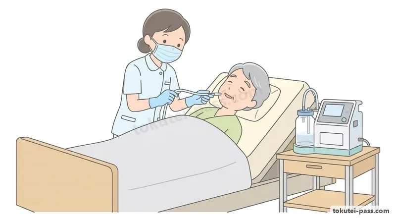
喀痰吸引を行う前には、まず医師の指示書を確認し、利用者に説明して同意を得たうえで、呼吸状態(呼吸音・ゴロゴロ音・SpO2・顔色・チアノーゼの有無)を観察し、安楽な姿勢(仰臥位または上半身を軽く起こした姿勢)を整えます。
Before suctioning sputum, verify the physician's instruction sheet, explain the procedure and obtain the user's consent, observe respiratory status (breath sounds, rattling sounds, SpO2, facial color, cyanosis), and position the user comfortably (supine or semi-reclined).
吸引器の準備では、吸引用チューブを折り曲げてスイッチを入れ、規定の「吸引圧(負圧/陰圧:成人の口腔・鼻腔ではおおむね20kPa[約150mmHg]以下、最大でも26kPa以下)」に調整されているかを確認します。
When preparing the suction machine, kink the suction tubing and turn on the switch to confirm that the Suction Pressure (negative pressure) is set to the prescribed range (for adult oral/nasal suctioning, generally 20 kPa / ~150 mmHg or less, maximum 26 kPa or less).
挿入する深さの絶対原則として、口腔内吸引では嘔吐反射や粘膜損傷を防ぐため「咽頭の手前まで(口蓋垂[のどちんこ]の裏側より奥へ入れない)」とし、気管カニューレ内部吸引では「カニューレの内部(先端を越えない深さ)」にとどめます。
As an absolute rule for insertion depth, oral suctioning must stop Just Before the Pharynx (do not insert past the uvula to prevent gag reflex and mucosal injury), and tracheal cannula suctioning must remain Inside the Cannula (not beyond the distal tip).
低酸素血症や粘膜損傷を防ぐため、1回の吸引時間は「15秒以内(気管カニューレ内は10〜15秒以内)」とし、カテーテルを指先でやさしく回しながら引き抜きます。
To prevent hypoxemia and mucosal damage, each suctioning pass must last Within 15 Seconds (10–15 seconds inside a tracheal cannula), gently rotating the catheter between the fingertips while withdrawing it.
また複数の部位を吸引する場合は、最も清潔度が高く感染に弱い部位から順に行うため、「①気管カニューレ内部 → ②鼻腔内 → ③口腔内」の順序を守り、気管カニューレ吸引に使ったカテーテルの再利用や、口腔・鼻腔用カテーテルを気管カニューレに入れることは絶対に厳禁です。
When suctioning multiple sites, proceed from the cleanest, most infection-vulnerable site first: (1) Inside the tracheal cannula -> (2) Nasal cavity -> (3) Oral cavity. Reusing a catheter after tracheal cannula suctioning or inserting an oral/nasal catheter into the tracheal cannula is strictly forbidden.
Key points
- 口腔内吸引は「咽頭の手前まで」、気管カニューレ内部吸引は「カニューレ先端を越えない範囲」まで挿入する
Insert the catheter only to Just Before the Pharynx for oral suctioning, and Within the Cannula Tip for tracheal cannula suctioning - 1回の吸引時間は低酸素防止のため「15秒以内」とし、吸引圧(陰圧)をおおむね20kPa以下に確認する
Each suctioning pass must be Within 15 Seconds to prevent hypoxia, with negative pressure verified at ~20 kPa or less - 吸引の順序は清潔度の高い「気管カニューレ内部 → 鼻腔内 → 口腔内」の順に行う
Suction in order from highest cleanliness: Inside tracheal cannula -> Nasal cavity -> Oral cavity
| Word | Meaning |
|---|---|
| 咽頭の手前まで(口腔内吸引) | Up to before the pharynx (oral suctioning depth) |
| 吸引圧(陰圧確認) | Suction pressure (negative pressure verification) |
| 吸引時間(1回15秒以内) | Suctioning duration (within 15 seconds per pass) |
| 無菌操作(気管カニューレ内吸引) | Aseptic technique (tracheal cannula suctioning) |
消化器系の解剖生理と経管栄養(胃ろう・腸ろう・経鼻)の種類
Digestive System Anatomy/Physiology and Types of Tube Feeding (Gastrostomy, Enterostomy, Nasogastric)
飲食物の通り道である消化管は、口腔 → 咽頭 → 食道 → 胃(入口の「噴門」から胃底部・胃体部を経て出口の「幽門」へ) → 小腸(十二指腸・空腸・回腸:栄養素の大半を消化吸収) → 大腸(水分吸収) → 肛門へと続きます。
The digestive tract runs from the Oral Cavity -> Pharynx -> Esophagus -> Stomach (entering at the Cardia, passing the fundus and body, exiting at the Pylorus) -> Small Intestine (duodenum, jejunum, ileum: digests and absorbs most nutrients) -> Large Intestine (absorbs water) -> Anus.
胃の入口にある「噴門(下部食道括約筋)」は胃内容物が食道へ逆流するのを防ぎ、出口の「幽門」は十二指腸へ送り出す量を調節しています。
The Cardia (lower esophageal sphincter) at the stomach entrance prevents stomach contents from refluxing into the esophagus, while the Pylorus at the exit regulates the amount emptied into the duodenum.
口腔から十分な食事が摂れないが消化管機能が保たれている場合に行う「経管栄養」には、鼻から胃までチューブを通す「経鼻経管栄養」、腹壁から胃に直接ろう孔をつくる「胃ろう(PEG)」、空腸等につくる「腸ろう」があります。
When oral intake is insufficient but gut function is preserved, Tube Feeding methods include Nasogastric Tube Feeding (tube from nose to stomach), Gastrostomy (PEG: fistula directly through the abdominal wall into the stomach), and Enterostomy (into the jejunum, etc.).
胃ろうカテーテルは、腹壁外の形状により「ボタン型(皮膚面から出っ張らず自己抜去しにくい・逆流防止弁あり)」と「チューブ型(接続が容易)」の2つがあり、胃内固定板の形により「バルーン型(滅菌蒸留水で膨らませる・1〜2か月ごとに交換)」と「バンパー型(4〜6か月ごとに交換)」に分かれます。
Gastrostomy catheters are classified by external shape into Button Type (flush with the skin, hard to self-remove, has an anti-reflux valve) and Tube Type (easy to connect), and by internal bumper shape into Balloon Type (inflated with sterile distilled water, replaced every 1–2 months) and Bumper Type (replaced every 4–6 months).
なお、胃ろう(PEG)を造設していても、嚥下機能に応じて口から食べる楽しみ(経口摂取)を併用することは可能であり、また接続チューブを外せば普段通りに湯船に浸かって「入浴」することもできます(ろう孔をガーゼで密閉保護する必要はありません)。
Even with a gastrostomy (PEG), users can still enjoy oral eating according to swallowing ability, and once the connecting tube is detached, they can Bathe in a bathtub as usual without sealing the stoma site with waterproof gauze.
Key points
- 胃の入口は「噴門(逆流防止)」、出口は「幽門」であり、栄養素は主に「小腸」で吸収される
The stomach entrance is the Cardia (prevents reflux) and the exit is the Pylorus; nutrients are mainly absorbed in the Small Intestine - 胃ろうカテーテルは体外形状で「ボタン型・チューブ型」、胃内固定板で「バルーン型・バンパー型」の4組合わせがある
Gastrostomy catheters have 4 combinations of external shape (Button/Tube) and internal bolster (Balloon/Bumper) - 胃ろう造設者でも状態に応じた経口摂取は可能であり、通常通りそのまま入浴することもできる
Users with a gastrostomy can still eat orally if swallowing permits and can bathe directly in a tub as usual
| Word | Meaning |
|---|---|
| 噴門と幽門 | Cardia and Pylorus |
| 胃ろう(PEG)・腸ろう・経鼻経管栄養 | Gastrostomy (PEG), Enterostomy, and Nasogastric tube feeding |
| ボタン型・チューブ型カテーテル | Button-type and Tube-type catheters |
| バルーン型・バンパー型 | Balloon-type and Bumper-type internal bolsters |
経管栄養の準備・体位・注入手順と口腔・皮膚のケア
Preparation, Positioning, Infusion Procedure, and Oral/Skin Care in Tube Feeding
経管栄養の実施前には、医師の指示書(栄養剤の種類・量・温度・滴下速度・時間)と利用者の氏名・体調(発熱・腹痛・下痢・便秘・腹部膨満感の有無)を確認し、排泄や吸引は栄養剤注入前に済ませておきます。
Before tube feeding, check the physician's instruction sheet (formula type, volume, temperature, drip rate, time) and the user's name and condition (fever, abdominal pain, diarrhea, constipation, abdominal distension); toileting and suctioning should be completed beforehand.
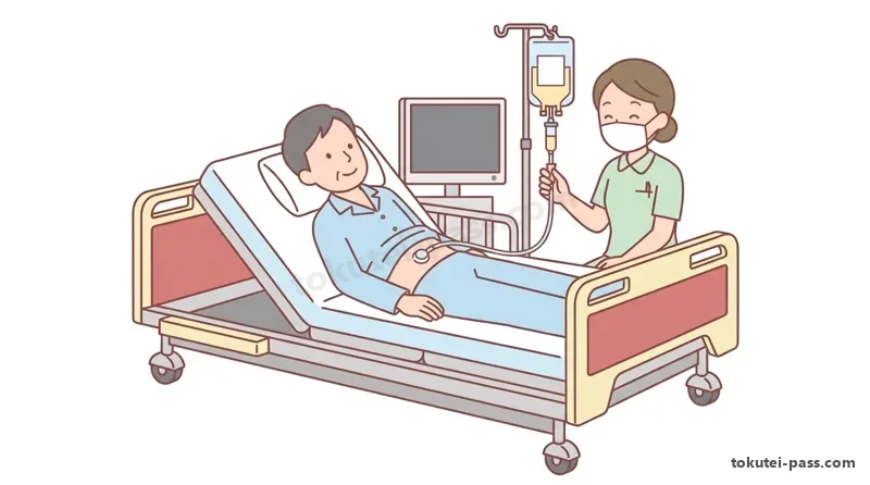
栄養剤の胃食道逆流による誤嚥性肺炎を防ぐため、注入時の体位はベッドの頭側を「30〜45度以上(または座位)」に挙上した「ファーラー位(半座位)」とし、注入終了後も「30分〜1時間程度」は同じ姿勢を保ちます。
To prevent aspiration pneumonia from gastroesophageal reflux, position the user in Fowler's Position (semi-sitting) with the head of the bed elevated 30–45 Degrees or More (or sitting), and maintain this posture for 30 to 60 minutes after infusion ends.
栄養剤は冷たすぎると下痢を起こしやすいため「人肌程度(約37〜38℃)」に湯煎等で温め(半固形化栄養剤や一部製品は常温のまま)、イリゲーター(栄養ボトル)とチューブ内を栄養剤で満たして空気を抜きます(クレンメを閉じる)。
Because cold formula easily causes diarrhea, warm liquid formula in a water bath to Body Temperature (~37–38°C) (semi-solid formulas and certain products are given at room temperature), prime the irrigator bottle and tubing with formula to expel air, and close the roller clamp (klemme).
胃ろう・腸ろう周囲の皮膚に発赤・腫脹・浸出液・不良肉芽やチューブの抜け・破損がないかを確認し、ストッパーが皮膚から1〜1.5cm程度の余裕がありスムーズに回転するかを見てから、指示された速度(クレンメ調整)で滴下を開始します。
Inspect the peristomal skin for redness, swelling, exudate, granulation tissue, or tube displacement, verify that the external bolster has 1–1.5 cm of play and rotates smoothly, and then start the drip at the prescribed rate using the roller clamp.
注入終了後はチューブの閉塞や細菌繁殖を防ぐため「白湯(微温湯)」を流してフラッシュします。また経管栄養中の利用者は咀嚼が減り唾液分泌が低下して口腔内細菌が増殖しやすいため、誤嚥性肺炎予防として毎日の「口腔ケア」が極めて重要です。
After infusion, flush the tube with Lukewarm Water to prevent occlusion and bacterial growth. Furthermore, because tube-fed users chew less and have reduced saliva secretion, oral bacteria multiply easily; daily Oral Care is vital to prevent aspiration pneumonia.
Key points
- 逆流・誤嚥防止のため、注入中および終了後30分〜1時間は「30〜45度以上の半座位(ファーラー位)」を保つ
Maintain a 30–45+ degree semi-sitting position (Fowler's position) during infusion and for 30–60 minutes afterward to prevent reflux and aspiration - 液体栄養剤は下痢防止のため「人肌程度(約37〜38℃)」とし、終了後は「白湯」を流して閉塞を防ぐ
Warm liquid formula to body temperature (~37–38°C) to prevent diarrhea, and flush the tube with lukewarm water after completion - 口から食べていなくても唾液減少により口腔内細菌が増えるため、毎日の「口腔ケア」を徹底する
Even without oral eating, reduced saliva increases oral bacteria, so thorough daily Oral Care is essential to prevent aspiration pneumonia
| Word | Meaning |
|---|---|
| ファーラー位(半座位:30〜45度以上) | Fowler's position (semi-sitting: 30–45 degrees or more) |
| イリゲーターとクレンメ | Irrigator container and Roller clamp (Klemme) |
| 半固形化栄養剤 | Semi-solid enteral formula |
| 白湯(微温湯)によるフラッシュ | Flushing with lukewarm water |
喀痰吸引・経管栄養における合併症・ヒヤリハットと緊急時対応
Complications, Near-Misses (Hiyari-Hatto), and Emergency Response in Suctioning and Tube Feeding
喀痰吸引中の異常・合併症として、吸引時間が長すぎることによる「低酸素状態(SpO2低下・チアノーゼ)」、粘膜損傷による「出血(血性痰)」、咽頭奥への刺激による「嘔吐反射」や迷走神経反射(徐脈・血圧低下)があります。
Complications during sputum suctioning include Hypoxia (drop in SpO2, cyanosis) from overly long suctioning, Bleeding (bloody sputum) from mucosal injury, Gag/Vomiting Reflex from deep pharyngeal stimulation, and Vagal Reflex (bradycardia, hypotension).
もし気管カニューレが抜けてしまった(事故抜去)場合や、吸引中に著しい出血・呼吸状態悪化が見られた場合は、介護福祉職が自分でカニューレを再挿入することはせず、直ちに看護職員や医師に連絡して指示を仰ぎます。
If a tracheal cannula accidentally dislodges or severe bleeding/respiratory deterioration occurs during suctioning, the care worker must never attempt to reinsert the cannula themselves, but immediately contact nursing staff or a physician for instructions.
経管栄養における主なトラブル・合併症には、栄養剤の注入速度が速すぎることや温度が低すぎることによる「下痢」、さらには栄養剤が急速に腸へ流れ込み冷や汗・めまい・動悸・低血糖などを起こす「ダンピング症候群(特に腸ろうで注意)」があります。
Major complications in tube feeding include Diarrhea caused by an overly rapid infusion rate or cold formula, and Dumping Syndrome (rapid entry of formula into the intestine causing cold sweats, dizziness, palpitations, and hypoglycemia—especially common in enterostomy).
また経鼻経管栄養では、咳き込みや体動によりチューブの先端が胃から食道や気管へずれてしまう危険があるため、注入前には看護職員による胃内留置の確認や口腔内でのチューブのとぐろ巻き(ループ)がないかを必ず確かめます。
In nasogastric tube feeding, coughing or movement can dislodge the tube tip from the stomach into the esophagus or trachea; before feeding, nursing staff must verify gastric placement and care workers must check that the tube is not coiled in the mouth.
経管栄養の注入中に利用者に「悪心(吐き気)・嘔吐・激しい咳き込み・顔色不良・呼吸苦」が現れた場合は、誤嚥や窒息を防ぐため「直ちにクレンメを閉じて注入を中止」し、顔を横に向けて口腔内を吸引・観察しつつ速やかに医師・看護職員へ報告します。
If the user develops Nausea, Vomiting, Severe Coughing/Choking, Pallor, or Dyspnea during tube feeding, Immediately Close the Clamp to Stop the Infusion to prevent aspiration or choking, turn the user's head to the side, suction the oral cavity as needed, and report to the physician or nurse.
Key points
- 経管栄養の注入速度が速すぎると「下痢」や「ダンピング症候群(冷や汗・めまい・動悸)」を起こしやすい
An overly rapid infusion rate easily causes Diarrhea or Dumping Syndrome (cold sweats, dizziness, palpitations) - 注入中に嘔気・嘔吐・咳き込みが見られたら、「直ちにクレンメを閉じて注入を中止」し医療職へ報告する
If nausea, vomiting, or coughing occurs during feeding, Immediately Close the Clamp to Stop the Infusion and report to medical staff - 気管カニューレや胃ろうカテーテルが抜けた場合、介護福祉職は再挿入せず直ちに医師・看護職員へ連絡する
If a tracheal cannula or gastrostomy catheter dislodges, do not reinsert it yourself; contact a physician or nurse immediately
| Word | Meaning |
|---|---|
| ダンピング症候群 | Dumping syndrome |
| 迷走神経反射 | Vagal reflex |
| 直ちに注入を中止(嘔吐・むせ時) | Immediately stop infusion (upon vomiting or choking) |
| カニューレ・カテーテルの事故抜去 | Accidental dislodgement of cannula/catheter |
Medical Care
Practice this chapter in the appOfficial references
- 公益財団法人社会福祉振興・試験センター「介護福祉士国家試験 試験科目別出題基準(医療的ケア)」 — https://www.sssc.or.jp/kaigo/kijun/index.html
- 厚生労働省「喀痰吸引等制度・介護職員等による喀痰吸引・経管栄養研修テキスト」 — https://www.mhlw.go.jp/stf/seisakunitsuite/bunya/hukushi_kaigo/seikatsuhogo/tannkyuuinn/index.html